Surgical Treatment of Racemose Cysticercosis in the Quadrigeminal Cistern: A Case Series
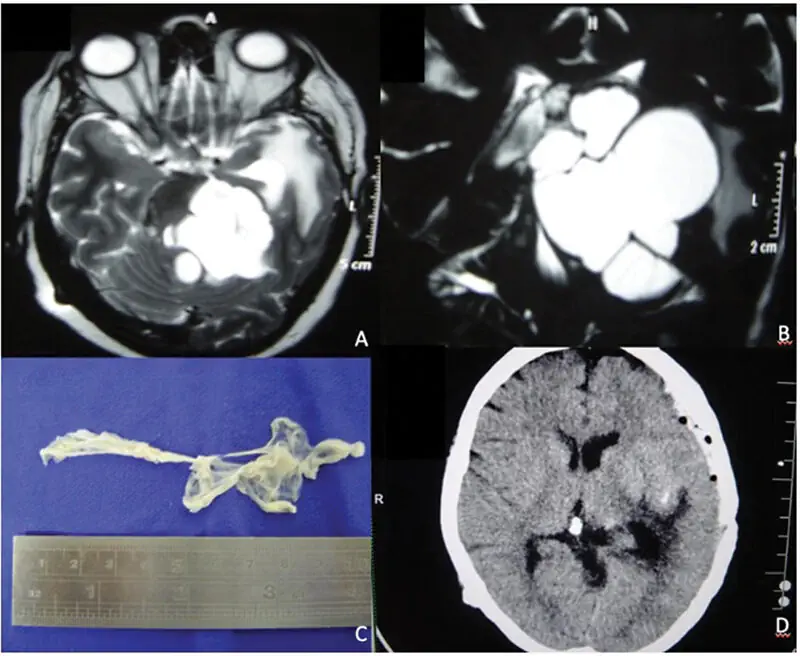
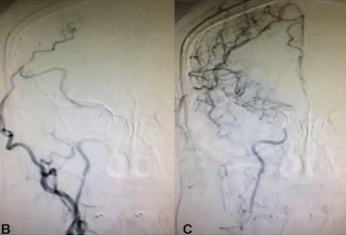
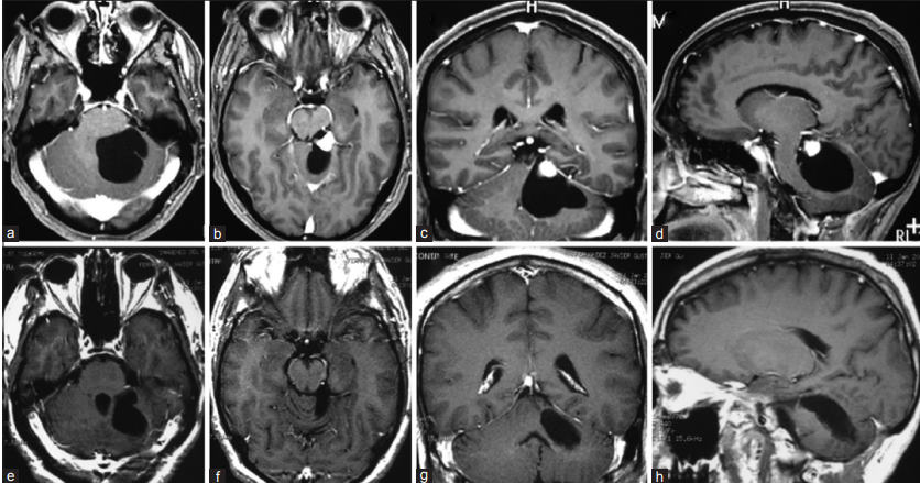
Introduction Racemose neurocysticercosis is rare and distinctive among the variety of neurocysticercosis pathologies, and it is characterized by the development of cysts in the basal subarachnoid region. This uncommon presentation involves the formation of multiple, non-encapsulated cystic membranes resembling a bunch of grapes due to the exogenous budding of aberrant proliferating Taenia solium larvae. Typically observed in expansive areas of the brain, such as the suprasellar, sylvian, and quadrigeminal cisterns, or around the rostral brainstem, these cysts lack scolex, do not always involve edema, and can escape detection with contrast enhancement depending on their life cycle. Case Description In both cases presented here, the patients’ multilobulated cysts had components in the posterior incisural space that extended below the mesial temporal region. Consequently, there was no need for a more complex approach to the pineal region, and cyst removal was achieved solely with the subtemporal approach. This is possible due to the cysts’ lack of adherence to neurovascular structures. Following cyst drainage, their capsules were easily removed with a forceps. Conclusion To achieve a successful outcome, these cases require a comprehensive understanding of neurocysticercosis variations as well as individualized surgical planning based on lesion characteristics and solid postoperative pharmacological management. The effective treatment of neurocysticercosis is clearly complex and still evolving, and continued vigilance and research will enhance our ability to manage the challenges this parasitic neurological condition presents.